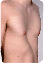
What is Pectus Excavatum?
Pectus excavatum is a disease in which the center of the chest collapses to form a funnel such as those used in natural science experiments and at home. It is believed that "costal (rib) cartilage grows faster than the sternum and ribs, so the middle of the sternum collapses because it cannot maintain a balance and bows to pressure from either side," but the pathogenesis is not yet clear. It may tend to be three to four times more prevalent among males than females. |
|||
Pectus excavatum is hereditary (domestic emergence); some children are born with it congenitally, while others develop it gradually. In childhood, it may accompany enlarged tonsils or hypertrophy of adenoids, and taking a deep breath due to the narrow pathway for air causes this collapse. Pectus excavatum may be improved by performing surgery for enlarged tonsils or hypertrophy of adenoids in advance. Many children who are diagnosed with Pectus excavatum are leptosome, having a flat chest and underdeveloped muscles and naturally tend to hunch over to breathe more easily, which leads to scoliosis (curvature of the spine) in many cases. The symptoms of diagnosed children indicates that breathing is compromised by the heart being pushed to the left, which may cause rapid heartbeats or breathlessness during exercise, as well as proneness to fatigue, frequent respiratory infections, and arrhythmia, because the collapse of Pectus excavatum is essentially progressive. |
 |
||
■ Medical treatment of Pectus Excavatum Methods of treatment can be divided into roughly three categories. |
|||
[1]Sternum Elevation Procedure *This is also called the Ravitch technique, named after Dr. Ravitch, who developed it. |
|||
*Costal cartilage ablation method (Sternocostal elevation technique) Although it has frequently been observed that the technique of largely ablating costal cartilage and rebuilding only part of the rib or costal cartilage restrains chest development, this technique is free from such anxiety. Re-collapse is also rare, because excessively long parts of the rib or costal cartilage are directly cut off. However, collapse can become highly visible due to excessively long ribs during adolescent growth spurts. Deformation of the chest may become apparent as height rapidly increases during adolescence, because patients with Pectus excavatum tend to be tall and slender, especially in cases of males. |
|||
[2]Sternal Rotation Procedure |
|||
[3]Chest Way Procedure |
|||
|
|||
Copyright © 2011 SOLVE Co., Ltd. All Rights Reserved.
